Otosclerosis
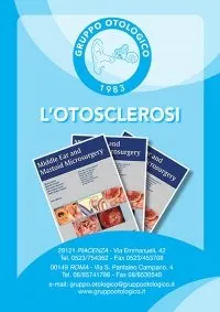
TEST 2 L’otosclerosi è una malattia dell’orecchio medio, non ereditaria ma che si presenta in ambito familiare anche a distanza di generazioni. È una malattia della staffa, un piccolo ossicino situato nell’orecchio medio che, in presenza di otosclerosi, non trasmette più correttamente il segnale sonoro alla coclea. L’otosclerosi è dovuta alla crescita di una massa ossea attorno alla staffa, che irrigidisce quest’ultima e ne blocca i movimenti (staffa sclerotica da “sclerosi”, termine che indica un indurimento e/o irrigidimento). Questa patologia determina una graduale perdita dell’udito (ipoacusia) e, se non curata, può degenerare in sordità completa.
La terapia attuale consiste nell’uso di protesi acustiche esterne e nella possibilità di operare chirurgicamente.
WHAT IS OTOSCLEROSIS?
Otosclerosis is also called otospongiosis, a term used in French-speaking countries. It is called otospongiosis because it describes the characteristics of the focus that causes the stapes to become fixed and thus leads to deafness. Otosclerosis is a disease present among populations of the Mediterranean basin, although the cause of this localization is unknown. Otosclerosis is not common in the Far East and Sub-Saharan Africa. There are different types of otosclerosis:
- stapedial otosclerosis, or “blockage” of the stapes without involvement of the cochlea;
- mixed otosclerosis, or stapedial “blockage” with involvement of the cochlea;
- cochlear otosclerosis with deafness due to a lesion of the cochlea.
WHEN DOES OTOSCLEROSIS OCCUR?
Otosclerosis occurs, in most cases, after the third decade of life (after the age of 30). It is very rare in younger individuals and extremely rare in childhood. It is also rare for it to occur after the age of 60. Otosclerosis is more common in women.
WHAT ARE THE SYMPTOMS OF OTOSCLEROSIS?
The symptoms of otosclerosis are varied:
- deafness: the most frequent symptom, which manifests as bilateral deafness (both ears) or unilateral deafness (deafness in one ear). The deafness caused by otosclerosis is of the conductive type in most cases (stapedial otosclerosis). Conductive deafness refers to the “blockage” of the ossicular chain (particularly the stapes).
Sometimes the deafness caused by otosclerosis is of the type:
- mixed (blockage of the stapes and cochlear involvement);
- sensorineural (not operable, but can be aided with a hearing aid or cochlear implant).
- tinnitus: tinnitus is a bothersome noise (commonly called “ringing in the ear”) that is not operable and often does not disappear after surgery. Its origins are unknown, and science has yet to find the causes or solutions to this disorder;
- vertigo or instability: this symptom has not yet been explained by science, although it is rare in cases of otosclerosis. Vertigo is the unpleasant sensation of not being physically balanced and can cause nausea.
HOW IS OTOSCLEROSIS DIAGNOSED?
Otosclerosis is diagnosed during a visit with an ENT specialist with:
- collection of symptoms (deafness and any tinnitus and vertigo) and transmission of family history;
- objective examination through an otoscope (viewing the external auditory canal and tympanic membrane) called otoscopy;
- audiometric test that determines the degree of deafness in one ear (unilateral) or both ears (bilateral). The audiometric test also studies the type of deafness (conductive, mixed, or sensorineural);
- impedancemetry with stapedial reflex study. This test examines the motility of the tympano-ossicular system and the presence or absence of the stapedial reflex (i.e., the mobility of the stapes that transmits sound) to further confirm the diagnosis of otosclerosis;
- speech discrimination: speech discrimination is the study of hearing quality (as opposed to the audiometric test which studies the quantity of hearing loss);
- vestibular test (rarely necessary) in which the type of vertigo or instability is studied if present as symptoms;
- radiological examination (CT scan without contrast) is rarely indicated. The radiological examination is used only in rare cases of sensorineural hearing loss to demonstrate possible cochlear involvement of the otosclerotic focus.
HOW IS OTOSCLEROSIS TREATED?
The true treatment for stapedial otosclerosis (i.e., the “blockage” of the stapes) with conductive hearing loss is surgery. The remedy for stapedial otosclerosis is surgery, performed under local anesthesia, without any external cuts, through the ear canal. Therefore, there will be no scars after the surgery. The ear with poorer hearing is operated on with a procedure called stapedotomy.
The other ear is operated on no sooner than 10-12 months later, to allow for the stabilization of the result of the first surgery. Even in mixed otosclerosis, the best therapy is surgery. The procedure is always stapedotomy. In this case, the hearing recovery is partial because the cochlear lesion is not recovered.
However, the recovery of residual hearing may allow the use of a hearing aid, with a satisfactory final hearing result. In cochlear otosclerosis
surgery is contraindicated, but in this case, it is possible to apply a hearing aid to improve hearing. In some rare cases of advanced cochlear otosclerosis (when the cochlea is ossified), where the use of a hearing aid is not possible, surgery can be performed to recover hearing with acochlear implant.
RISKS AND COMPLICATIONS OF STAPEDOTOMY FOR OTOSCLEROSIS
The major risks associated with stapedotomy involve the preservation of hearing.
In most cases (97%), the patient fully recovers hearing after the surgery.
In 2% of cases, hearing is only partially recovered. In 1% of cases, hearing is not recovered or, very rarely, it may worsen or be lost. Other risks of stapedotomy include:
- perforation of the tympanic membrane (which can be repaired during the same surgery);
- post-operative infection, which requires antibiotic therapy;
- rarely, facial nerve paralysis may occur a few days later. In this case, it is resolved with anti-inflammatory therapy within a few weeks;
- even rarer is facial nerve paralysis after the surgery.
FOLLOW UP: POST-OPERATIVE CHECK-UPS
The patient is discharged the same day or the next morning, in Day hospital.
After the stapedotomy, the patient will undergo antibiotic therapy for a week.
The post-operative check-up will be performed approximately 30 days after the surgery.
The check-up will involve:
- the removal of the gelatin inserted in the external auditory canal;
- a tonal audiometric test to ascertain hearing recovery;
- a vocal audiometric test to confirm the quality of hearing recovery.
The patient can, only after the first follow-up visit, wet the ear (take a shower, etc.)
A further check-up will take place after 6 months, and another one after a year from the surgery.
At this point, that is, a year after the surgery, the operation on the second ear can be planned.
THE EXPERIENCE OF THE OTOLOGICAL GROUP IN THE TREATMENT OF OTOSCLEROSIS
The Otological Group has 50 years of experience in the treatment of otosclerosis. It is the Italian center with the most cases of otosclerosis treated. Over 7,500 cases of otosclerosis have been operated on since 1975.
The Otological Group has published two textbooks on the subject, which form the basis of international study for any ENT specialist who wants to approach this challenging surgery.
The books on otosclerosis by the Otological Group are also the reference basis for national and international medico-legal disputes.
video
Doctors
Dott. Antonio Caruso
Otorinolaringoiatria, Otologia, Neurotologia, Chirurgia della Base Cranica, Chirurgia Endoscopica dei Seni Paranasali
Dott.ssa Vittoria Di Rubbo
Otorinolaringoiatria, Otologia, Chirurgia della Base Cranica, Neurotologia e Chirurgia Endoscopica dei Seni Paranasali
Dott. Giuseppe Fancello
Otorinolaringoiatria, Otologia, Neurotologia, Chirurgia della Base Cranica, Chirurgia Endoscopica dei Seni Paranasali, Laringologia
Dott.ssa Anna Lisa Giannuzzi
Otorinolaringoiatria, Otologia, Neurotologia, Chirurgia della Base Cranica, Vestibologia
Dott. Lorenzo Lauda
Otorinolaringoiatria, Otologia, Neurotologia, Chirurgia della Base Cranica, Chirurgia Endoscopica dei Seni Paranasali, Chirurgia Riabilitativa del Nervo facciale
Dott. Enrico Piccirillo
Otorinolaringoiatria, Neurotologia, Chirurgia della Base Cranica, Chirurgia Endoscopica dei Seni Paranasali, Oncologia Testa-Collo
Dott. Gianluca Piras
Otorinolaringoiatria, Otologia, Neurotologia, Chirurgia della Base Cranica, Chirurgia Endoscopica dei Seni Paranasali
Dott.ssa Alessandra Russo
Otorinolaringoiatria, Otologia, Neurotologia, Chirurgia della Base Cranica, Chirurgia Ricostruttiva del Padiglione Auricolare
Prof. Mario Sanna
Otorinolaringoiatria, Otologia, Neurotologia, Chirurgia della Base Cranica
Dott. Abdelkader Taibah
Otorinolaringoiatria, Otologia, Neurotologia, Chirurgia della Base Cranica
contacts
Via Morigi, 41
29122, Piacenza (PC)
Tel. (+39) 0523.751280
WhatsApp – 389.2625175
ufficio.privati@casadicura.pc.it

