Paraganglioma
Paraganglioma, or glomal tumour, is a benign tumour that originates from certain corpuscles responsible for controlling blood pressure distributed along the course of certain nerves in the middle ear and skull base.
WHAT IS PARAGANGLIOMA?
It is a rare neuroendocrine tumour that can develop in various regions of the body (including the head, neck, chest and abdomen). Approximately 97% of cases are benign and can be removed by surgery.
Although it is still called Glomic Tumour or Chemodectoma, the term Paraganglioma is the currently accepted and preferred one. Three types of lesion can be defined:
- the Glomus Tympanicus and Jugularis;
- Carotid Paraganglioma (or Carotid Glomus Tumour);
- the Vagal Paraganglioma.
Paragangliomas are richly vascularised whose size and location can lead to various symptoms, from pulsating tinnitus to hearing loss to functional alterations of the cranial nerves. Depending on the place of origin of the Paragangliomas and subsequent growth, they are distinguished into the following types:
- Class A paraganglioma – Tumour limited to the tympanic cavity;
- Paraganglioma classe B – Tumore esteso all’ipotimpano e alla mastoide
- Paraganglioma class C – Tumour involving the skull base.
Paraganglioma therapy is surgical.
WHERE DOES PARAGANGLIOMA ARISE AND HOW DOES IT DEVELOP?
Based on location, paragangliomas are subdivided according to a classification, which has very important therapeutic implications.
- Class A – tympanic paraganglioma (limited to the tympanic cavity);
- Class B – tympanomastoid paraganglioma (extension to the hypotympanum and mastoid);
- Class C – tympanic-jugular paraganglioma (skull base involvement);
- C1: extension limited to the vertical section of the internal carotid artery;
- C2: Knee-limited extension of the internal carotid artery;
- C3: extension limited to the horizontal section of the internal carotid artery;
- C4: involvement of the anterior foramen lacerum area;
- If class C tumours extend intracranially this extension can be classified into De (extradural) and Di (intradural);
There are two other types of paragangliomas, the cervical paragangliomas:
- vagal paraganglioma: located in the neck at the level of the vagal nerve;
- carotid paraganglioma: alized in the neck at the level of the carotid bifurcation.
WHAT ARE THE SYMPTOMS OF PARAGANGLIOMA?
Because of their rich vascularisation (the entire tumour mass is filled with blood) these tumours give rise to a quite specific disorder: pulsating tinnitus (pulsating noise in the ear).
Naturally, the size and location of the tumour determine the presence of other symptoms, first and foremost hearing loss.
In cases of extensive paragangliomas (class C to be precise), it is possible, albeit rarely, to notice symptoms such as dysphonia (altered timbre of voice), dysphagia (difficulty swallowing), inability to lift the shoulder, inability to move the tongue and a paralysis of the half-volvement on the affected side.
HOW IS PARAGANGLIOMA DIAGNOSED?
In most cases, the diagnosis is clinical.
A red, pulsating retrotimpanic mass is evident on otoscopy (often synchronous with heartbeats), occupying portions (particularly the inferior portion) or the entirety of the tympanic area.
It is absolutely contraindicated to perform a diagnostic biopsy either on an outpatient basis or in the operating theatre because of the risk of high bleeding. It will in fact be the subsequent radiological examinations that will confirm the diagnostic suspicion. High-resolution CT scans of the temporal bone and skull base allow, in the case of class A or B paragangliomas, confirmation of extension to the eardrum case or mastoid. In the case of class C paraganglioma, this radiological examination allows the degree of involvement of the intrapetrosal tract (i.e. inside the temporal bone, in which the ear is contained) of the internal carotid artery to be assessed. This is crucial for planning both the most appropriate surgical intervention and for securing the internal carotid artery (a vital structure) prior to removal of the paraganglioma. MRI of the brain and skull base with paramagnetic contrast medium (gadolinium) allows further diagnostic confirmation and, above all, differentiation from other neoplasms of the skull base.
Paraganglioma, due to its intense internal vascularisation, presents a typical ‘salt and pepper’ signal, which allows it to be distinguished from other lesions (e.g. neurinomas or meningiomas of the jugular foramen).
Also in the case of larger paragangliomas (class C), other complementary examinations (arteriography and CT angiography or MRI angiography) make it possible to quantify the vascularisation and understand the relationship with vital structures.
WHAT IS THE THERAPEUTIC MANAGEMENT OF PARAGANGLIOMA?
Treatment is closely related to the classification, as well as to the age and symptoms reported by the patient. Tympanic paraganglioma (class A): This is a small paraganglioma located exclusively in the tympanic cavity, visible otoscopically as a reddish retrotympanic mass.
Depending on its size, it is removed through the external ear canal, or it may require a retroauricular incision. In both cases, the tympanic membrane must be lifted to gain access to the tympanic cavity. The operation requires 1 night’s hospital stay.
Paraganglioma tympano-mastoid (class B): In addition to involving the tympanic case, it extends to the hypotympanum and mastoid.
Its removal always requires a retroauricular incision. It may also be necessary to remove the ossicles and perform the operation in two stages (the second for reconstruction of the ossicular chain).
Sometimes it is necessary to cut down the posterior wall of the ear canal and then perform an open tympanoplasty, resulting in an enlargement of the canal.
In particularly extensive cases, it may be necessary to completely remove the outer and middle ear, obliterating the residual cavity with abdominal fat and closing the external ear canal like a sack.
The operation is almost always performed under general anaesthesia and requires 2-3 days’ hospitalisation.
The tympanic-jugular paraganglioma (class C) needs a separate chapter.
WHAT IS THE MANAGEMENT OF JUGULAR PARAGANGLIOMA (class C)?
Jugular tympanic paraganglioma (class C) is an important tumour because of its location at the level of the jugular foramen. The latter is a bony foramen located in an area of the skull base rich in important nerve and vascular structures.
More precisely, they transit there:
- the glossopharyngeal nerve that contributes to the function of swallowing;
- the vagus nerve which has multiple functions, the most important of which are those of vocal cord movement (phonation) and swallowing;
- the accessory nerve responsible for part of the lifting movement of the shoulder;
- the bulb of the jugular vein, which drains most of the blood from the middle of the head.
Since the jugular foramen is located immediately inferior to the middle ear, tumour growth within the tympanic cavity is very common, resulting in symptoms such as hearing loss and pulsating tinnitus.
The presence of the tumour in the case almost always makes it visible during the otoscopic examination.
In the course of their extension, these tumours may come to affect other structures, such as the internal carotid artery, which is the most important artery carrying blood to the brain, the hypoglossal nerve, which allows the movement of half the tongue, the inner ear in its auditory and balance (vestibular) components, and the facial nerve, which controls the movement of the muscles of half the face.
Involvement of other nerves, such as those involved in eye motility and the vertebral artery, is rarer.
The involvement of various nerves by the tumour does not mean that these nerves do not function; on the contrary, their pre-operative function is often normal.
Usually the paraganglioma passes through the jugular foramen and thus consists of an intracranial part and an extracranial part (in the neck). When the tumour extends internally to the dura (the outermost layer of the meninges) and thus comes into contact with the brain structures, it may be necessary to divide the operation into two separate surgical stages, to be performed about 6 months apart. This is to reduce the postoperative risk of a leakage of the fluid surrounding the brain (liquorrhoea) and consequently the risk of possible meningitis.
PRE-OPERATIVE EVALUATION
Due to the complexity of this type of tumour, a correct evaluation requires a thorough otoneurological examination supplemented by audiological tests, and in particular an accurate radiological study using high-resolution CT and MRI. The two examinations are complementary in that the former allows an accurate assessment of the bone erosion, while the latter through the infusion of a paramagnetic contrast medium allows the tumour extension to be visualised at the intracranial level and in the neck. PRE-OPERATIVE TREATMENT The rich vascularity of the tumour requires some special pre-operative measures. The first two are routinely performed in all patients with tympanic-jugular paraganglioma; the others are reserved only for paragangliomas with particular extension.
1. Arteriography. This consists of a radiological study of the vascular system carried out under local anaesthesia by introducing an iodinated contrast medium through an artery in the thigh (femoral artery). Arteriography gives very important information on the nature of the tumour, its vascularisation, its relationship with neighbouring structures, and makes it possible to identify exactly which arteries carry blood to the tumour. The examination also makes it possible to study the veins draining blood from the head.
In particular cases, the main venous outlet is the bulb of the jugular vein on the side to be operated on. Since it is not possible to spare this vessel during surgery, in such a situation it is advisable to postpone the operation in order to avoid problems due to cerebral venous stasis. Generally, tumour growth leads to a slow closure of the bulb of the jugular vein with simultaneous compensatory development of secondary vessels, which makes it possible to perform the operation.
2. Embolisation. This procedure closes the vessels that carry blood to the paraganglioma by placing small metal particles or coils inside the vessels. It is performed under general anaesthesia by inserting a thin probe through the femoral artery into the affected vessels.
For this treatment to be most effective, it must be carried out in the 72 hours preceding surgery and requires hospitalisation. 3. Stent reinforcement. Some tumour extensions carry a significant risk of accidental rupture of the carotid artery during surgery. In order to reduce these risks, it may be decided to reinforce the wall of the affected section of the carotid artery by introducing a Stent. This is also a procedure that is carried out via the femoral artery and should be performed approximately 40 days before the planned operation. 4. Balloon occlusion. When the carotid artery is particularly at risk, detachable balloons are placed inside the affected artery during surgery in order to permanently occlude it. This procedure should be performed at least 30 days before surgery, so that the brain can get used to the new blood supply situation. Not all patients are able to tolerate this procedure, which must always be preceded by an occlusion test. The latter consists of introducing a small probe with a balloon at the end through the femoral artery. This balloon is inflated once the correct position inside the affected artery has been reached. For about ½ h, the patient’s neurological state is then monitored to assess whether the blood supply from the other vessels is sufficient to supply the cerebral structures.
SURGICAL TREATMENT
The surgical procedure most frequently used in the removal of this type of paraganglioma is called infratemporal route type A. The incision starts above the ear and extends to the neck. During the operation, the facial nerve, even when not affected by the tumour, has to be moved from its bony channel in order to have better access to the area affected by the tumour. This displacement results in a temporary, more or less complete deficit of nerve function, with good functional recovery within a few months in most cases. When the facial nerve is invaded by the tumour, it is necessary to remove the involved part and then reconstruct it with a graft of another nerve (usually taken from an ankle). In this case, recovery of half-face function is protracted and always partial. Hearing, which is almost always compromised by the tumour itself, is never recoverable, as removal of the tympanic membrane and ossicular system with sack-like closure of the external auditory canal is necessary. Often, due to the tumour extension, it is also necessary to sacrifice the inner ear, resulting in total deafness on the operated side. Saving the nerves involved during tumour removal surgery is particularly difficult. When the nerves involved are sacrificed, there are problems in the postoperative period, particularly related to swallowing difficulties, with the risk that food may end up in the lungs causing pneumonia. These difficulties are generally overcome quickly in young patients (within 5-6 days), with more difficulty in the elderly, so much so that in some cases it is preferable to perform partial tumour removal or simple clinical and radiological follow-up rather than risk damaging the nerves. Nerve paralysis already present before surgery as caused by the paraganglioma makes the post-operative period easier. This is because the paraganglioma generally causes gradual paralysis, which is much better compensated for by the body than sudden paralysis, which frequently occurs after surgery. In some cases, the tumour invades a part of the bone (occipital condyle) that is responsible for stabilising the head. When it is necessary to remove this bone during surgery, a collar is used post-operatively as a precautionary measure. In the event of permanent head instability, a subsequent operation to fix the head to the cervical spine is indicated (a very rare occurrence). In cases of major intradural invasion, this component is left in place to be removed during a second surgery. The latter is performed 3-4 months later via an access similar to the first operation, but without the extension to the neck. Staging significantly reduces the risk of a liquorrhoea in the neck, which is difficult to control. At the end of the operation, the surgical cavity is filled with fat taken from the abdomen.
ALTERNATIVE TREATMENT OF PARAGANGLIOMA
Tympanic-jugular paragangliomas, although insidious, are slow-growing benign tumours. In cases of normal function of the lower cranial nerves (i.e. absence of dysphonia and dysphagia with normal laryngeal motility) and in elderly patients (> 65-70 anni), simple radiological follow-up may be advisable. Radiological follow-up is performed once a year with CT and MRI or more simply with an MRI with contrast medium. As far as the use of radiotherapy is concerned, this is indicated in elderly patients or in the case of tumour residues at the level of the cavernous sinus (these are surgical residues of tympanic-jugular paraganglioma extending beyond the anterior foramen lacerum, the surgical removal of which would entail, in addition to the risk of bleeding, a lesion of the nerves regulating ocular motility). Radiotherapy treatment is not curative, but by damaging the vessels that bring blood to the tumour, it generally tends to stop its growth for a few years. A surgical exeresis following radiotherapy treatment can be very complex. In addition, recent molecular studies carried out by our group are showing that paraganglioma has biological features that confer radioresistance (i.e. after an initial halt to tumour growth due to necrosis of the peripheral tumour tissue, a radiotherapy-induced stimulation of tumour growth would occur).
THE CAROTID PARAGANGLIOMA
Carotid paraganglioma originates in the neck at the level of the carotid bifurcation. It normally manifests itself as an asymptomatic swelling, sometimes producing a cranial nerve deficit. Other times, the presence of this type of paraganglioma is diagnosed during angiographic studies of patients with multiple paragangliomas. The main radiological feature, due to its site of origin, is the enlargement of the carotid bifurcation, which is visible on both CT and MRI scans and particularly in angiographic studies. Surgical treatment is the main therapeutic modality and is carried out through a neck-only approach. Of course, the technical difficulties are mainly related to the size of the tumour and the consequent involvement of the carotids. In more complex cases, pre-operative treatment by insertion of a carotid stent may be necessary, as described in the section on tympanic-jugular paragangliomas.
WHAT ARE THE RISKS OF SURGERY?
The differentiation between tympanic paraganglioma and tympano-mastoid paraganglioma must also be made with regard to the risks of surgery. Surgery to remove class A and B paragangliomas falls under middle ear surgery with risks similar to those of a tympanoplasty, such as:
- Infection
- Worsening of hearing
- Vertigo
- Perforation of the tympanic membrane
Extremely rare is the possibility of more serious complications such as leakage of cerebral fluid, meningitis, brain abscess and surgical damage to the facial nerve resulting in paralysis of half the face. With regard to paraganglioma type C, the main surgical risks are:
- Worsening of deafness/total deafness on the operated side;
- paralysis/temporary paralysis of the face;
- dysphagia (difficulty in feeding);
- dysphonia (decreased tone of voice);
- difficulty in lifting the shoulder;
- paralysis of half the tongue;
- ab ingestis’ pneumonia;
- liquorrhoea (leakage of the fluid in which the brain is immersed).
In the case of a lesion with significant involvement of the internal carotid artery or with intradural extension, very low percentages may occur:
- death;
- major neurological deficits such as temporary or permanent hemiplegia (paralysis of half the body).
CARE OF THE PARAGANGLIOMA PATIENT
Assuming a normal post-operative course and in experienced hands, the patient operated on for paraganglioma type C is cared for in the first 18-24 hours post-operatively in an intensive/sub-intensive care unit. The clinical evaluation, followed by a post-operative encephalic CT scan on the day after surgery, makes it possible to verify the absence of immediate complications (in particular ongoing bleeding or complications affecting the encephalic structures) and the subsequent transfer to the ordinary in-patient ward. Approximately one week of hospitalisation will follow. In the case of paralysis of the nerves responsible for swallowing and voice, in order to reduce the risk of pneumonia, it is recommended to eat semi-solid food (fruit smoothies, crème caramel, etc.) until a good degree of compensation has been achieved. The patient will in any case begin a logopaedic rehabilitation process during hospitalisation to normalise both phonatory and swallowing function. Rehabilitation treatment will be continued at home until compensation is achieved. In the event of failure to recover, a small operation on the larynx (injection of fat or artificial material at the level of the vocal cord) can improve the clinical condition. No weights should be lifted for about two months after surgery to reduce the possibility of late onset liquorrhoea. In the case of facial paralysis, the eye should be protected with artificial tears, ointments, special eyeglasses or eyelid weights and periodic eye examinations until the eyelid closes again. Electrical stimulation of the paralysed muscles is not recommended, as such a procedure leads to muscle fibrosis with permanent sequelae. From the moment the patient feels the onset of motility recovery, it may be useful to perform movements in front of the mirror, trying to maintain facial symmetry. In the case of vertigo in the post-operative phase, early mobilisation accelerates vestibular compensation. In order to complete this compensation, the patient is asked to repeat the movements that bother him/her the most.
THE IMPORTANCE OF A CENTRE OF EXCELLENCE FOR THE TREATMENT OF PARAGANGLIOMA
What has been described above confirms that surgery for auditory nerve neurinoma requires experience and the highest degree of specialisation. Only teams with oto-neurosurgical expertise and a system of clinical, radiological and rehabilitation care can guarantee the patient optimal results while minimising the possibility of post-surgical complications. The Otology Group, active since 1983, has contributed over the years to developing innovative surgical techniques that have made it possible to reduce not only mortality, but also the morbidity of the operation (by morbidity we mean the incidence of serious and disabling post-operative complications for the patient). With around 700 paragangliomas surgically treated over these years and more than 70 cases undergoing simple radiological monitoring, the Otology Group represents one of the world’s reference centres for the treatment and management of this pathology, with the highest number of cases in Europe and estimated at international level. As proof of this, in September 2023, Prof. Mario Sanna’s Otology Group organised the first international congress on the rare disease; it was a unique event with the active participation of specialists in the field from more than 25 different countries around the world.
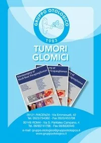
Microsurgery of Skull Base Paragangliomas book
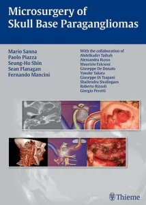
This is the Otology Group’s latest book, recently presented at the 2nd European Academy of ENT and Head and Neck Surgery (EAORL-HNS), held in Nice in April 2013.
This book offers a comprehensive overview of paraganglioma, from the analysis of pathophysiological and clinical aspects to a detailed description of all surgical procedures, with rich photographic documentation.
Read the review HERE
The book has also been translated into Chinese.
Doctors
Dott. Antonio Caruso
Otorinolaringoiatria, Otologia, Neurotologia, Chirurgia della Base Cranica, Chirurgia Endoscopica dei Seni Paranasali
Dott.ssa Vittoria Di Rubbo
Otorinolaringoiatria, Otologia, Chirurgia della Base Cranica, Neurotologia e Chirurgia Endoscopica dei Seni Paranasali
Dott. Giuseppe Fancello
Otorinolaringoiatria, Otologia, Neurotologia, Chirurgia della Base Cranica, Chirurgia Endoscopica dei Seni Paranasali, Laringologia
Dott.ssa Anna Lisa Giannuzzi
Otorinolaringoiatria, Otologia, Neurotologia, Chirurgia della Base Cranica, Vestibologia
Dott. Lorenzo Lauda
Otorinolaringoiatria, Otologia, Neurotologia, Chirurgia della Base Cranica, Chirurgia Endoscopica dei Seni Paranasali, Chirurgia Riabilitativa del Nervo facciale
Dott. Enrico Piccirillo
Otorinolaringoiatria, Neurotologia, Chirurgia della Base Cranica, Chirurgia Endoscopica dei Seni Paranasali, Oncologia Testa-Collo
Dott. Gianluca Piras
Otorinolaringoiatria, Otologia, Neurotologia, Chirurgia della Base Cranica, Chirurgia Endoscopica dei Seni Paranasali
Dott.ssa Alessandra Russo
Otorinolaringoiatria, Otologia, Neurotologia, Chirurgia della Base Cranica, Chirurgia Ricostruttiva del Padiglione Auricolare
Prof. Mario Sanna
Otorinolaringoiatria, Otologia, Neurotologia, Chirurgia della Base Cranica
Dott. Abdelkader Taibah
Otorinolaringoiatria, Otologia, Neurotologia, Chirurgia della Base Cranica
contacts
Via Morigi, 41
29122, Piacenza (PC)
Tel. (+39) 0523.751280
WhatsApp – 389.2625175
ufficio.privati@casadicura.pc.it

