Neuroma
The acoustic nerve neurinoma is a benign tumor that affects the eighth cranial nerve and is one of the most frequent intracranial tumors.
This neoplasm originates from the sheath that covers the eighth nerve itself.
The acoustic nerve neurinoma, also known as vestibular schwannoma as it originates from Schwann cells, is a benign form of brain tumor originating from Schwann cells with symptoms that include unilateral sensorineural hearing loss.
What is an acoustic neurinoma?
The acoustic nerve neurinoma is a benign tumor that affects the eighth cranial nerve.
The eighth cranial nerve is composed of two branches: the cochlear branch (essential for hearing) and the vestibular branch (essential for maintaining balance).
Symptoms vary depending on the size of the tumor and the involvement of surrounding nerve structures.
The neurinoma can grow to considerable sizes and may affect nearby cranial nerves or the brainstem.
Where does the acoustic neurinoma originate and how does it develop?
The VIII cranial nerve (or vestibulocochlear nerve) has two components: an auditory part (cochlear nerve) and a vestibular part (vestibular nerve), closely related to the facial nerve (which controls facial movement).
The neurinoma can originate and develop either in the internal auditory canal (the bony channel of the petrous ridge where these nerves run) or in the cerebellopontine angle. As the neurinoma grows, it expands towards the structures of the central nervous system (the brainstem), causing a functional alteration of this structure. Due to its slow growth, the body gradually adapts, alternating with periods of more or less evident symptoms.
What are the symptoms of acoustic nerve neurinoma?
This brain tumor, also called vestibular schwannoma due to its location, although benign, compresses the acoustic nerve and surrounding structures, causing disabling disorders. Therefore, the acoustic neurinoma is almost never asymptomatic and the symptoms vary depending on the size of the tumor and the compression of the surrounding nerve structures.
The vast majority of patients typically exhibit sensorineural hearing loss on the affected side (i.e., involving the acoustic nerve), which can be progressive and of varying degrees or, less frequently, sudden or fluctuating.
In an initial phase, the associated symptoms can include various hearing disorders, sometimes persistent, such as unilateral hearing loss, tinnitus (ringing), vertigo, and difficulty maintaining balance.
Acute vertigo attacks are not frequent, although a general sense of disequilibrium or instability is reported in almost all cases.
The growth of the tumor can compress other nerves and tissues, causing coordination problems, unilateral weakness of the facial muscles, headaches, facial paresis, and hydrocephalus, which is the blockage of cerebrospinal fluid circulation.
When the compression affects the trigeminal nerve, symptoms can include, in addition to hearing symptoms, facial sensitivity disorders.
If the tumor also compresses the facial nerve, there may be taste disturbances and facial paresis.
The severity of symptoms is directly proportional to the size of the neurinoma.
If the neuroma reaches a size greater than 25 mm in the cerebellopontine angle, it tends to compress the trigeminal nerve, and therefore, in association with auditory symptoms, facial sensitivity disorders with hypoesthesia or hyposensitivity of the face may appear. In cases of even larger tumors, a slight facial nerve deficit (mild paresis), taste disorders, but rarely a complete nerve paralysis, may occur. Diplopia (double vision) or intracranial hypertension (headache with nausea and vomiting) with papilledema and urinary incontinence are symptoms of large or giant neuromas (diameter in the cerebellopontine angle > 4 cm).
When the tumor is of considerable size (diameter over 3-4 cm) and compresses the brainstem, various symptoms may arise such as:
- loss of muscle coordination;
- double vision (diplopia);
- intracranial hypertension (which causes headache with nausea and vomiting).
Diagnosis and causes of acoustic neuroma
The diagnosis of acoustic neuroma is not simple and immediate. The symptoms, in fact, present themselves gradually, at the pace of the tumor’s growth, which occurs slowly (on average 1-2 millimeters per year) and without a precise progression.
In the initial phase, the first signs of the pathology can be confused with those of other disorders such as labyrinthitis or Menière’s disease.
The essential diagnostic tests are magnetic resonance imaging (MRI) and computed tomography (CT), both with contrast medium.
The audiometric test and some neurological tests are also necessary to quantify the hearing loss and verify the functionality of the facial muscles.
A simple audiometric test, performed in the clinic, generally reveals unilateral sensorineural hearing loss.
The deafness is associated with a decrease in word discrimination in the examined ear. Another very sensitive, inexpensive, and simple test, the ABR or auditory brainstem response study, allows us to detect early signs of compression and stretching of the cochlear nerve indicative of the possible presence of the tumor.
It is now common clinical practice, in the presence of unilateral sensorineural hearing loss, to refer the patient for a brain MRI with paramagnetic contrast medium (gadolinium). With this technique, tumors of a few millimeters can be diagnosed and, in cases of large tumors, the extent and relationships with cerebral vessels and neurological structures of the cerebellopontine angle can be evaluated.
The CT scan is sometimes required to study the anatomical characteristics of the temporal bone and for this reason, it should be considered a complementary test to the MRI. Knowledge of the tumor’s relationships with neurovascular structures allows for planning the most suitable surgical access route, taking into account the hearing status, age, and general condition of the patient, as well as the hearing in the opposite ear. It is of fundamental importance, through an accurate diagnosis, to outline the characteristics of an acoustic neuroma to set the most appropriate therapy, surgical and non-surgical.
Treatment of acoustic nerve neuroma
The acoustic neuroma can be treated in various ways, and the therapy is closely related to three factors: tumor size and location, symptoms, and the patient’s age/health condition.
- In the case of an intrameatal acoustic nerve neurinoma (i.e., confined within the internal auditory canal), a wait-and-scan policy is normally adopted, meaning the patient undergoes regular magnetic resonance imaging checks (once a year). If there is growth or the appearance of symptoms (e.g., vertigo), surgery may be indicated.
The surgery to remove the neurinoma is performed under general anesthesia, using an operating microscope. If hearing is normal, speech discrimination is good, and evoked potentials are normal or with very slight alterations, the surgical approach used is the middle cranial fossa. If the tumor invades the medial part without reaching the bottom of the internal auditory canal, with good hearing and good speech discrimination, the retrosigmoid approach combined with a retrolabyrinthine approach is indicated. In cases with compromised hearing or socially unusable hearing, even in the presence of intrameatal tumors, the translabyrinthine approach is used as in larger tumors. It is worth remembering that, in the case of an elderly patient (>65-70 years) and the presence of vestibular-type symptoms , intratympanic injection of gentamicin may be indicated from a therapeutic point of view. This antibiotic, given its toxicity to the cells of the balance organ, results in an improvement in most cases of acute vertigo, without subjecting the patient to the risks associated with surgery. - In the case of a small acoustic nerve neurinoma (i.e., protruding from the internal auditory canal and extending < 1 cm into the cerebellopontine angle), the therapeutic policy is similar to the previous case. If surgery is indicated, if the size of the neurinoma does not exceed 0.5 cm outside the canal and hearing and speech discrimination are good, the surgical approach used is the middle cranial fossa. If the neurinoma is slightly larger, but still less than 1 cm and does not reach the bottom of the internal auditory canal, with good hearing and speech discrimination, it will be removed via the retrosigmoid approach combined with a retrolabyrinthine approach.
In cases with compromised or socially unusable hearing, even in the presence of small tumors, the translabyrinthine approach is used as in larger tumors.
In selected cases (intrameatal or small neurinomas), it is possible to rehabilitate hearing function simultaneously with the removal of the lesion by placing a cochlear implant, always through the translabyrinthine approach. The cochlear implant is a fully implantable prosthesis capable of restoring hearing by directly stimulating the cochlear nerve. Its application, therefore, requires the complete anatomical and functional integrity of the cochlear nerve after the removal of the neurinoma.
- A medium-sized acoustic nerve neurinoma (1.0 – 2.0 cm) extends from the internal auditory canal to the cerebellopontine angle, without causing compression on the brainstem and brain. The surgery to remove a neurinoma of this size is performed via an extended translabyrinthine approach, making an incision behind the ear, above the mastoid bone.
The mastoid and inner ear structures are removed to expose the tumor. It is then completely removed without manipulating the cerebellum. Occasionally, partial removal may be performed (especially to preserve the anatomical continuity of the facial nerve). The lack of mastoid bone is filled with abdominal fat. The translabyrinthine approach sacrifices the hearing and balance mechanism. Consequently, the ear becomes permanently deaf. Although the balance mechanism has been removed from the operated ear, the contralateral vestibular apparatus generally provides stability to the patient within a period ranging from 1 to 4 months. - A large acoustic nerve neurinoma (2.0 – 4.0 cm) is large enough to cause pressure on the brainstem and disturb its vital centers.
The procedures for large tumors require a wider bone removal to adequately expose the neoplasm and control the large blood vessels that obstruct access to the tumor itself. For this reason, special vascularization investigations (arteriography and magnetic resonance angiography) may be required. The surgical approach (extended translabyrinthine route) is always performed through an incision behind the ear, above the mastoid bone. The mastoid, the structures of the inner ear, and a portion of cranial bone are removed to expose the neoplasm. The tumor is then completely removed unless intraoperative vital alterations occur. If there are changes in blood pressure values, pulse rate, or breathing, the surgical act may be interrupted before the tumor is completely removed (in this case, a second intervention is necessary to complete the tumor removal). However, this occurrence is extremely rare. The lack of mastoid bone is filled with fat taken from the abdomen.
- In giant neuromas (greater than 4 cm), the percentage of complications is higher, especially in elderly patients where partial removal is preferred to reduce compression at the brainstem level. For non-elderly patients, the strategy is the same as for large neuromas. In the presence of intracranial hypertension, the insertion of a cerebrospinal fluid drainage shunt may be indicated before the removal of the neuroma. The surgical route for the removal of the neoplasm is either translabyrinthine or transotic.
What is the role of radiotherapy in the treatment of acoustic nerve neuroma?
Radiotherapy is indicated for small tumors in elderly patients or in cases of tumors in the only hearing ear.
It is a technique based on the principle that a high dose of radiation concentrated on a small area can stop tumor growth without interfering with the functioning of surrounding tissues.
The radiation source can come from either radioactive cobalt (Gamma Knife) or a linear accelerator (LINAC).
This treatment can stop tumor growth for a certain period, but it does not make the mass disappear nor cause a reduction in volume.
In some cases, despite radiotherapy, the tumor continues to grow. The surgical removal of an irradiated tumor that continues to grow can be more complex and risky (especially due to an increased likelihood of facial nerve paralysis) than that of a non-irradiated tumor. The Gamma Knife is now proposed by many centers as a therapeutic alternative to surgery.
However, it is a relatively recent method and easily accepted by patients as it is non-invasive, but before scientifically confirming the validity of this treatment, a longer-term follow-up of treated cases is needed. It should also be noted that the Gamma Knife is not a risk-free treatment, and numerous complications are reported in the literature, such as hearing loss in 30% of treated cases, facial paralysis or paresis in 20%, trigeminal neuralgia in 10%, and cerebral edema plus intracranial hypertension in 10-15%.
Recently, cases of malignant transformation of the tumor following radiotherapy have been reported worldwide.
For these reasons, in our opinion, microsurgical removal remains the elective treatment for acoustic tumors.
In centers with established experience in operations with tumors of the same size, the results of microsurgery are comparable if not superior and safer than radiotherapy.
What are the risks of the surgical intervention?
Some of the main and possible risks associated with acoustic nerve neuroma surgery are:
- Hearing loss: in small tumors, it is sometimes possible to save hearing by removing the tumor. However, the majority of neoplasms are large, and hearing in the affected ear is lost following surgical removal. The patient can continue to lead a completely normal life with only one hearing ear;
- Tinnitus: in most cases, the noise remains unchanged, as before the surgery. In 10% of cases, it may be more intense after the surgery, and in some cases (about 20-30%), the noise may also disappear;
- Taste disturbances and dry mouth: taste disturbances and dry mouth are common for a few weeks after surgery. In 5% of patients, these disturbances may last longer;
- Vertigo and balance disorders: in acoustic nerve tumor surgery, it is also necessary to remove part or all of the balance nerve. In most cases, it is also necessary to remove the labyrinth. Since the balance nerve is often damaged by the tumor, its removal almost always results in an improvement in preoperative instability.
In the post-operative phase it is common to experience vertigo-related disorders, even in acute phases, and symptoms may last for days or weeks.
Imbalance or instability with head movement may persist for a long time (in 30% of patients), until compensation by other balance centers occurs.
- Facial nerve paralysis: the acoustic nerve neurinoma can be in close contact with the facial nerve (the nerve that controls the movement of the muscles responsible for closing the eye and those that control facial expression).
Following the removal of an acoustic nerve tumor, temporary paralysis of the affected side of the face may occur; this results in facial asymmetry with inability to close the eye.
The facial nerve paralysis can be the result of edema, direct nerve injury, or compromised blood supply.
Only through careful surgery with the aid of a microscope and intraoperative monitoring of the facial nerve is it possible to preserve the integrity and functionality of the nerve. Despite this, transient facial paralysis may still occur.
Generally, recovery occurs spontaneously. The recovery of facial functionality varies greatly from person to person; it can occur within a few weeks or several months (up to 10-12 months) and may not be 100% complete.
Beyond this period, the chances of recovery are very low and the surgeon may propose a second surgery to connect the facial nerve to a branch of the V cranial nerve (masseter-facial anastomosis) to prevent muscle atrophy in the face and allow the eye to close. In 5% of cases, the facial nerve is completely enveloped or incorporated by the tumor mass. In very rare cases, the tumor may also originate directly from the facial nerve (facial nerve neurinoma).
In both of these conditions, it is necessary to sever the facial nerve to remove the tumor.
In such cases, it may be possible to immediately reconstruct the nerve using a sural nerve graft (taken from the leg).
If this is not possible, a subsequent surgery can be performed to connect the facial nerve to the masseter nerve.
- Ocular complications: facial nerve paralysis often makes the eye dry and defenseless. Consultation with an ophthalmologist may be indicated, as it may be necessary to apply artificial tears, use a protective eyepatch and/or an external eyelid weight or partial surgical eyelid closure to reduce the risk of keratitis or corneal ulcer.
These measures keep the eye moist, thus avoiding ocular complications. - Decreased functionality of other nerves in large tumors: the acoustic nerve neurinoma can come into contact with the nerves that control the ocular muscles, face, mouth, and throat. These nerves can be injured and cause double vision, numbness of the throat, face, and tongue, shoulder mobility weakness, dysphonia, and difficulty swallowing.
These problems can remain permanently, although this is very rare. - Brain complications and death: acoustic nerve tumors are located near vital brain centers that control breathing, blood pressure, and heart function.
Since the neuroma has a progressive growth, it can come into contact with these brain centers and can engulf blood vessels that supply these areas of the brain. Careful and meticulous dissection of the tumor generally allows avoiding such complications. If the blood supply to these brain centers is compromised, serious complications can occur: loss of muscle control, paralysis, and even death.
In our experience, perioperative mortality is less than 1% in small and medium tumors; this percentage slightly increases in large or giant tumors (2-3%).
- Post-operative cerebrospinal fluid loss and meningitis: surgery for acoustic nerve neuroma involves a temporary loss of cerebrospinal fluid (the fluid that surrounds the brain). To avoid this complication after tumor removal, the cavity is filled with fat taken from the abdomen. Occasionally, fluid leakage may occur after the surgery, and to stop it, it may be necessary to reoperate or insert a lumbar drain. Immediate control and resolution of the fluid leakage are crucial to prevent the onset of meningitis, which is a serious infection of the fluid and tissue around the brain. In the presence of this complication, prolonged hospitalization is required.
It is often indicated, moreover, to treat with high doses of antibiotics.
- Post-operative hemorrhage and cerebral edema: hemorrhage and cerebral edema can develop after surgery for acoustic nerve tumor (1%). If it occurs, an emergency intervention may be necessary to open the wound, stop the bleeding, and allow the brain to re-expand.
Patient care after acoustic neuroma surgery
In the case of a normal post-operative course and in experienced hands, the patient operated on for neuroma is assisted in the first 18-24 hours post-operatively in intensive/sub-intensive care. Clinical evaluation followed by a post-operative brain CT scan the day after the surgery allows verifying the absence of immediate complications (particularly ongoing bleeding or complications affecting brain structures) and the subsequent transfer to the ordinary ward.
This will be followed by about a week of hospitalization during which the patient will begin motor rehabilitation, also with the help of specialized physiotherapists in assisting this particular clinical condition. Upon discharge, the patient will return home able to maintain autonomous walking and the ability to independently perform normal daily activities.
The post-operative check-ups will be scheduled at intervals of 2 months, 6 months, and one year post-operatively.
In case of persistence of vestibular symptoms (particularly instability and difficulty maintaining balance during rapid head movements), the patient will continue the vestibular rehabilitation already started during the hospital stay at home.
In case of facial nerve paralysis, it will be necessary to wait at least the first 8-10 months to evaluate any therapeutic or rehabilitative measures to improve facial function, enhancing the ability to completely close the eyelid.
In the meantime, it is advisable for the patient to undergo periodic ophthalmological check-ups to monitor the condition of the cornea.
The rare cases with impaired function of the lower cranial nerves (responsible for swallowing and vocal cord movement) will continue with speech therapy, already started during the hospital stay.
It is important to remember that this latter situation is rarely encountered and only in cases of large or giant acoustic neuroma.
The same applies to cases with post-operative cerebellar ataxia; this condition involves instability and motor incoordination related to alterations and manipulation of the cerebellum. Thanks to the use of the translabyrinthine surgical approach, this clinical condition rarely manifests and is limited to cases of particularly extensive neuroma. As previously described, rehabilitative motor physiotherapy, already initiated during hospitalization, is continued upon discharge at home or possibly at a dedicated facility (such as the Vestibolar at Casa di Cura Piacenza as a Center of Excellence for the diagnosis, vestibular rehabilitation, and treatment of all balance-related pathologies, the first in Italy with a revolutionary evidence-based approach).
The importance of a Center of Excellence for the treatment of acoustic nerve neuroma
What has been described previously confirms that acoustic nerve neuroma surgery requires experience and high specialization.
Only teams with oto-neurosurgical skills and a system of clinical, radiological, and rehabilitative assistance can guarantee optimal results for the patient, minimizing the possibility of post-surgical complications.
The Gruppo Otologico, active since 1983, has over the years contributed to developing innovative surgical techniques that have reduced not only mortality but also the morbidity of the intervention (morbidity refers to the incidence of serious and disabling post-operative complications for the patient).
With the highest case volume in Italy of 3,800 surgeries for acoustic neuroma, the Gruppo Otologico represents one of the worldwide reference centers for the diagnosis, care, and treatment of the pathology. According to 2021 data, the National Outcomes Plan (PNE) of the Ministry of Health ranks it first in Italy for the number of middle ear surgeries and cochlear implants performed by the Gruppo Otologico di Piacenza.
Doctors
Dott. Antonio Caruso
Otorinolaringoiatria, Otologia, Neurotologia, Chirurgia della Base Cranica, Chirurgia Endoscopica dei Seni Paranasali
Dott.ssa Vittoria Di Rubbo
Otorinolaringoiatria, Otologia, Chirurgia della Base Cranica, Neurotologia e Chirurgia Endoscopica dei Seni Paranasali
Dott.ssa Anna Lisa Giannuzzi
Otorinolaringoiatria, Otologia, Neurotologia, Chirurgia della Base Cranica, Vestibologia
Dott. Lorenzo Lauda
Otorinolaringoiatria, Otologia, Neurotologia, Chirurgia della Base Cranica, Chirurgia Endoscopica dei Seni Paranasali, Chirurgia Riabilitativa del Nervo facciale
Dott. Enrico Piccirillo
Otorinolaringoiatria, Neurotologia, Chirurgia della Base Cranica, Chirurgia Endoscopica dei Seni Paranasali, Oncologia Testa-Collo
Dott. Gianluca Piras
Otorinolaringoiatria, Otologia, Neurotologia, Chirurgia della Base Cranica, Chirurgia Endoscopica dei Seni Paranasali
Dott.ssa Alessandra Russo
Otorinolaringoiatria, Otologia, Neurotologia, Chirurgia della Base Cranica, Chirurgia Ricostruttiva del Padiglione Auricolare
Prof. Mario Sanna
Otorinolaringoiatria, Otologia, Neurotologia, Chirurgia della Base Cranica
Dott. Abdelkader Taibah
Otorinolaringoiatria, Otologia, Neurotologia, Chirurgia della Base Cranica
News
Cochlear implant: when and what it is used for
Rely on a Health Center of Excellence for cochlear implant surgery is crucial for the patient, who will be guided through the multidisciplinary rehabilitation pathway for effective hearing recovery.
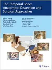
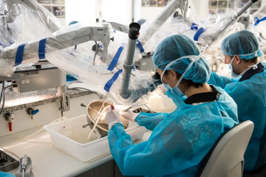
The Temporal Bone: Anatomical Dissection and Surgical Approaches
Temporal bone anatomy is arguably the most complex
NATIONAL & INTERNATIONAL TRAINING PROGRAMMES
NATIONAL & INTERNATIONAL TRAINING PROGRAMMES Practical Courses in Middle
Microsurgical Management of Middle Ear and Petrous Bone Cholesteatoma
The cholesteatoma, strictly speaking a cyst and not
contacts
Via Morigi, 41
29122, Piacenza (PC)
Tel. (+39) 0523.751280
WhatsApp – 389.2625175
ufficio.privati@casadicura.pc.it

