Cochlear Implant
The cochlear implant is a device that allows hearing for adults and children affected by profound deafness. It provides electrical impulses directly to the fibers of the auditory nerve bypassing the damaged cells of the inner ear (hair cells). Once the impulses reach the brain, they are interpreted as sounds. Cochlear implants are not hearing aids; they do not amplify sounds (as hearing aids do), but capture sound, convert it into signals/electrical impulses (as a cochlea would), and transfer the newly generated signals/electrical impulses to the cochlear nerve stimulating it. This stimulation of the cochlear nerve ensures the human perception and recognition of sound.
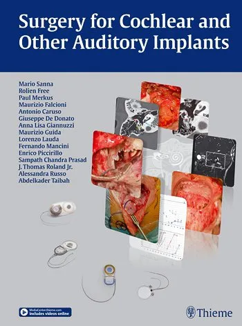
The cochlear implant consists of an internal component made up of the receiver/stimulator with the electrode array, which is surgically inserted into the ear, and an external component made up of the speech processor and the coil, which is usually applied behind the ear (retroauricular region).
The cochlear implant is indicated for subjects with bilateral profound deafness, either genetic or acquired during life, for whom hearing aids do not provide adequate help in sound perception. In certain subjects, the cochlear implant can significantly improve the quality of life (e.g., social, communicative, learning abilities) by enhancing the ability to hear words without the aid of visual signals (e.g., lip reading). Improving the recognition of normal everyday environmental sounds increases the ability to listen in a noisy environment and enhances the possibility of hearing during television programs, music, and phone conversations. The cochlear implant can be applied to those subjects who have severe or profound sensorineural hearing loss (anacusis) bilaterally, with little or no benefit from using a hearing aid and with strong motivation and expectations. The cochlear implant can be applied to both adults and children. A distinction must be made between prelingual and postlingual deafness:
- prelingual refers to before the acquisition of the ability to produce and understand language and speech;
- postlingual refers to after acquiring the ability to produce and understand language and speech.
In 60-70% of children, deaf from birth and implanted in the first three years of life, there is normal schooling, this percentage drops to 20-25% if implanted after the 3rd year of life.
Even children who are hearing-impaired from birth and have postlingual deafness can obtain very good results from a cochlear implant; the earlier the implantation procedure is carried out with respect to the onset of deafness, the better the results can be obtained.
Adults with postlingual deafness who have lost their hearing as a result of meningitis, trauma, otosclerosis, or other conditions with progressive hearing loss over time can benefit tremendously from cochlear implantation.
The concept, as with children, of early intervention also applies to adults.
Adults with prelingual deafness undergoing cochlear implantation may reap very limited benefits (sound discrimination).
Although many people are not aware of it, in Italy cochlear implantation is totally free of charge as it is paid for by the National Health System, and so is the rehabilitation process and the pathway to its activation. Financial assistance from the State can help resolve the discomfort caused by severe forms of sensorineural hearing loss by finding the most suitable solution.
A generic cochlear implant captures sounds from the external environment, converts the captured sounds into electrical impulses/signals, and finally transmits the generated electrical impulses/signals to the cochlear nerve, stimulating it.
The stimulation of the cochlear nerve ensures the perception and recognition of sounds.
L’intervento chirurgico di “inserzione di impianto cocleare” viene eseguito da uno specialista in otorinolaringoiatria in anestesia generale ed ha una durata di 2-3 ore.
Durante l’intero intervento viene eseguito il monitoraggio del nervo facciale. Viene praticata un’incisione della cute a forma di L rovesciata.
Una volta scollati i tessuti ed esposto l’osso si effettua una piccola mastoidectomia. Si esegue la timpanotomia posteriore, che ci permette di accedere alla regione della finestra rotonda. A questo punto si crea l’alloggiamento per il ricevitore-stimolatore con l’ausilio di un modellino metallico fornito nel kit dell’impianto. Si esegue, quindi, l’apertura della coclea (cocleostomia) e si inserisce (nella scala timpanica) il filo porta elettrodi che viene poi fissato con del tessuto connettivo più eventuale colla. Eseguito ciò, l’audiologo confermerà attraverso opportuni tests la corretta collocazione.
Occorre, inoltre, valutare eventuali interferenze tra gli elettrodi del filo ed il nervo facciale. Infine si esegue un’attenta chiusura dei lembi.
Nei casi in cui sia necessario impiantare un orecchio affetto da otiti croniche o precedentemente sottoposto a tecniche aperte è opportuno rimuovere tutta la patologia ed eseguire la chiusura del condotto uditivo esterno a cul di sacco con obliterazione della cavità al fine di evitare possibili complicanze (meningite). In presenza di infezioni attive, l’inserzione dell’impianto cocleare è posticipata ad un secondo tempo.
Nei pazienti affetti da malformazioni congenite è opportuno eseguire un’attenta valutazione radiologica attraverso una TC ed una RM ponendo particolare attenzione alla pervietà della coclea ed al decorso del nervo facciale. Nei casi di reimpianto bisogna porre particolare attenzione alla fibrosi cicatriziale a livello della timpanotomia posteriore e della finestra rotonda ed inoltre al lembo cutaneo.
Before the installation of a cochlear implant, the patient must undergo a series of tests to determine suitability for the procedure.
During these tests, the patient will also receive all the information regarding the installation procedure (what it involves, pre-operative guidelines, post-operative course, etc.). Before deciding to apply a cochlear implant, it is advisable to perform assessments:
- Audiological tests (e.g., pure tone audiometry and speech audiometry with and without hearing aids, tympanometry, auditory evoked potentials, promontory test)
- Clinical tests (routine blood tests for general anesthesia)
- CT and MRI (for evaluating the anatomy of the inner ear)
- Consultation and speech therapy visit
Today, thanks to the continuous advances in medical technology in the audiological field, the use of a cochlear implant represents an excellent remedy for resolving many cases of profound deafness and moderate to severe hearing loss.
Unfortunately, in Italy, there is still little knowledge and awareness of the positive impact on quality of life that the installation of a cochlear implant can bring.
In patients with a cochlear implant, there is an improvement in hearing ability and an awareness of sounds in daily life.
Generally, after the activation of the cochlear implant, patients can understand speech without lip reading and even talk on the phone. Learning to hear with a cochlear implant requires time, practice, determination, and patience.
On the day of activation, many patients can only hear ‘beeps’ or unrecognizable sounds, while others may already be able to understand voices. Fortunately, a great effort by the patient can be rewarded, as cochlear implants can significantly restore the ability to hear all sounds.
The installation of a cochlear implant is a procedure that carries some risks and can result in various complications. Among the possible risks and complications following the installation of a cochlear implant are:
- Infections: the surgical wound could become infected if not properly disinfected, but this is among the common complications of all types of surgery. It is essential not to wet the wound with water for a period specified by the doctor;
- Suffering/necrosis of the musculo-cutaneous flap: a musculo-cutaneous flap that is too thin could go into ischemia and, therefore, necrosis. In such a case, a surgical revision may be necessary;
- Extrusion of the receiver: this can be a consequence of the suffering/necrosis of the musculo-cutaneous flap and will require a revision surgery;
- Defective functioning of the receiver: a revision surgery will be necessary to replace it;
- Episodes of imbalance and tinnitus: a buzzing (tinnitus) is often present in patients with hearing loss.
After surgery, the buzzing generally decreases; occasionally, however, it may arise after the surgery; - Vertigo: temporary vertigo, usually lasting a few days, can occur in 5% of our patients; very rarely, this disturbance persists over time;
- Facial nerve paralysis: a possible but rare postoperative complication is temporary facial paralysis which generally recovers spontaneously within a few weeks;
- taste disturbances: the taste of the anterior third of the tongue on the operated side is ensured by a nerve that passes through the middle ear, it is often necessary to cut this nerve and this can cause a temporary taste disturbance in about 10% of patients.
This disturbance can last up to a year and only in rare cases remains permanent; - cerebrospinal fluid leak/meningitis: the loss of cerebrospinal fluid (cerebrospinal fluid leak) can occur from the opening of the cochlea. To avoid this problem, the area where the electrode enters the cochlea is sealed with fascia and fibrin glue. If this complication occurs in the postoperative period, a revision surgery will be necessary to prevent meningitis.
Meningitis, in very rare cases, can also occur as a consequence of the spread of an infection to the inner ear: in this case, the therapy consists of administering massive doses of antibiotics.
Il ricovero ha in media una durata di 3 giorni, trascorsi i quali il paziente ritorna a casa, facendo attenzione a non bagnare la ferita.
La rimozione dei punti della sutura avviene dopo 12-14 giorni dall’intervento e dopo circa 4 settimane il paziente dovrà tornare per l’attivazione dell’impianto cocleare e il relativo “mappaggio”, durante il quale saranno regolati gli elettrodi presenti nel filo porta elettrodi e sarà reso possibile sentire i suoni. Nel percorso riabilitativo il paziente, nel corso dei mesi successivi, dovrà tornare nuovamente per riprogrammare l’elaboratore al fine di ottimizzare la resa e per effettuare delle consulenze logopediche, con figure altamente specializzate nelle patologie del linguaggio, in grado di educare i pazienti alla corretta interpretazione dei suoni captati ed elaborati dall’impianto cocleare. I pazienti impiantati potranno condurre un tipo di vita normale e potranno effettuare, senza alcun disagio, tutte le consuete attività quotidiane. I pazienti impiantati potranno praticare sport evitando, se possibile, quelli che comportano scontri violenti (es. pugilato, arti marziali etc.) e utilizzare baschetti protettivi per altri tipi di sport più energici. Prima di entrare in acqua sarà necessario rimuovere i componenti esterni che non sono impermeabili, in modo da evitare la compromissione del loro effettivo utilizzo.
Relying on a Center of Excellence like the Gruppo Otologico means being able to count, within a specialized healthcare context, on a multidisciplinary team (otolaryngologists, audiometrists, audiologists, speech therapists, etc.) and on modern equipment and audiometric booths. The network of skills, implemented by the collaboration of the multispecialty team, allows us to accompany the patient through the entire rehabilitation process achieving the maximum results, thanks to the continuous interaction between the operators.
FAQ – COCHLEAR IMPLANT
The cost of a cochlear implant is significant, however, many do not know that in Italy the cochlear implant is completely free as it is covered by the National Health Service, as is the rehabilitation process and the path to its activation. The financial support from the State can help solve, by finding the most suitable solution, the discomfort caused by severe forms of sensorineural hearing loss.
The cochlear implant can be applied to those who have a severe or profound sensorineural hearing loss (anacusis) bilaterally, present from birth or acquired during life, with little or no benefit from the use of a hearing aid and with strong motivations and expectations.
The cochlear implant consists of an internal component made up of the receiver/stimulator with the electrode carrier wire (array), which is to be surgically inserted inside the ear, and an external component made up of the speech processor and the coil which is generally applied behind the ear (retroauricular region).
In patients with a cochlear implant, there is an improvement in hearing ability and an awareness of everyday life sounds. Every single sound heard with a cochlear implant passes through the small array of electrodes implanted in the cochlea. Generally, after the activation of the cochlear implant, patients can understand speech without lip-reading and even talk on the phone. Learning to hear with a cochlear implant requires time, practice, determination, and above all, patience. On the day of activation, many patients can only hear “beeps” or unrecognizable sounds, while others may already be able to understand voices. Fortunately, a great effort by the patient can be rewarded, as cochlear implants can effectively restore the ability to hear all sounds.
Before the installation of a cochlear implant, the patient must undergo a series of tests to determine eligibility for the procedure. Before deciding whether to apply a cochlear implant, it is appropriate to perform the following assessments:
- audiological exams (e.g., tonal audiometry and speech audiometry with and without hearing aids, tympanometry, auditory evoked potentials, promontory test)
- clinical exams (routine blood tests for general anesthesia)
- CT and MRI (to evaluate the anatomy of the inner ear)
- consultation and speech therapy visit
The installation of a cochlear implant is a procedure that carries some risks and can result in various complications. Among the different possible risks and complications following the installation of a cochlear implant are: infections, suffering/necrosis of the muscle-skin flap, malfunctioning of the receiver, moments of imbalance and tinnitus, dizziness, taste disturbances, cerebrospinal fluid leakage/meningitis.
In the post-operative period, the sutures are removed 12-14 days after the surgery, and the patient must return to the clinic after about 4 weeks for the activation of the implant and the related “mapping,” during which the electrodes in the electrode array are adjusted to enable hearing sounds. In the rehabilitation process, the patient must return to the clinic in the following months to reprogram the processor to optimize performance and to undergo speech therapy consultations with highly specialized professionals in language pathologies, who can educate patients on the correct interpretation of the sounds captured and processed by the cochlear implant.
contacts
Via Morigi, 41
29122, Piacenza (PC)
– Phone (+39) 0523.754362
– WhatsApp (+39) 378.3025085
– Email segreteria@gruppootologico.com
– www.gruppootologico.com
HOURS 08:30 – 17:00 from Monday to Friday
Doctors
Dott. Antonio Caruso
Otorinolaringoiatria, Otologia, Neurotologia, Chirurgia della Base Cranica, Chirurgia Endoscopica dei Seni Paranasali
Dott.ssa Vittoria Di Rubbo
Otorinolaringoiatria, Otologia, Chirurgia della Base Cranica, Neurotologia e Chirurgia Endoscopica dei Seni Paranasali
Dott. Giuseppe Fancello
Otorinolaringoiatria, Otologia, Neurotologia, Chirurgia della Base Cranica, Chirurgia Endoscopica dei Seni Paranasali, Laringologia
Dott.ssa Anna Lisa Giannuzzi
Otorinolaringoiatria, Otologia, Neurotologia, Chirurgia della Base Cranica, Vestibologia
Dott. Lorenzo Lauda
Otorinolaringoiatria, Otologia, Neurotologia, Chirurgia della Base Cranica, Chirurgia Endoscopica dei Seni Paranasali, Chirurgia Riabilitativa del Nervo facciale
Dott. Enrico Piccirillo
Otorinolaringoiatria, Neurotologia, Chirurgia della Base Cranica, Chirurgia Endoscopica dei Seni Paranasali, Oncologia Testa-Collo
Dott. Gianluca Piras
Otorinolaringoiatria, Otologia, Neurotologia, Chirurgia della Base Cranica, Chirurgia Endoscopica dei Seni Paranasali
Dott.ssa Alessandra Russo
Otorinolaringoiatria, Otologia, Neurotologia, Chirurgia della Base Cranica, Chirurgia Ricostruttiva del Padiglione Auricolare
Prof. Mario Sanna
Otorinolaringoiatria, Otologia, Neurotologia, Chirurgia della Base Cranica
Dott. Abdelkader Taibah
Otorinolaringoiatria, Otologia, Neurotologia, Chirurgia della Base Cranica
contacts
Via Morigi, 41
29122, Piacenza (PC)
Tel. (+39) 0523.751280
WhatsApp – 389.2625175
ufficio.privati@casadicura.pc.it

